Thermography & Head – Neck Care
This Photo by Unknown Author is licensed under CC BY-SA
Thermal Imaging: An Exciting Progression in the Treatment of Head & Neck Care
Thermal imaging offers a risk-free, non-invasive, relatively cost-effective, portable, and clinically tested means of early detection for a whole host of head & neck dysfunctions and diseases. As a result, it has offered potentially life-changing outcomes in patient care, quality of life, and mortality for countless patients.
Read on to find out why it might just offer an excellent choice in safeguarding the health of you and your family.
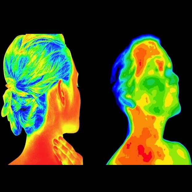
What is thermography?
Simply put, thermal imaging identifies picks up on anomalies in localized blood tissue temperature and blood flow. These anomolies can indicate anything from a minor injury, a benign growth, or an aggressive melanoma via the generation of a thermal image. These images clearly highlight changes in patterns of localized body heat that may need to be investigated further. It is exactly this unusual, localized pattern of body heat and blood flow that can alert a clinician to a whole host of potential health conditions.
Early Detection of Tumors
The growth of a tumor, for example, would require angiogenesis (new blood cell generation), greater metabolic activity (due to nutrient and growth needs), and greater blood flow (enhanced vascular density). All of that activity would push the body out of its delicate thermal homeostasis (balance, or equilibrium) in the localized area where the tumor is growing.
Thermal imaging could subsequently and immediately pick up on that temperature differential. This image would alert a clinician that something anomalous is happening in that area of your body. The identification of that something could save lives. It can vastly increase a patients’ survival outcomes and quality of life. And it could also, crucially, remove the need for aggressive later-stage treatments (such as chemotherapy) where the disease is to be diagnosed later.
In this way, thermal imaging screening offers a fantastic tool for early detection. Early detection means the opportunity to engage with more orthodox diagnostic tools earlier (e.g., MRI’s). This efficiency and medical value have earned thermography FDA recognition as an effective adjunct scanning tool for cancer and a whole host of other conditions.
Thermography: Advancing Head & Neck Care
One specific area of the body for which thermography – a test that relies on heat – has proven powerful is in the head & neck region, partially because ‘the human body is considered to be a stairway of heat with the highest temperature in the forehead and cervical regions’.
Subsequently, the efficacy of thermal imaging techniques in the early treatment of cerebrovascular disease, oral inflammation, thyroid dysfunction, periodontal disease, face & mouth cancer (including cervical lymph node metastasis and oral cancer) has emerged with strength in recent years.
Let’s take a closer look!
Cerebrovascular Disease
Cerebrovascular disease (CD) refers to a wide range of conditions that can cause a cerebrovascular event owing to a disruption or blockage of blood flow to the brain (e.g., deep vein thrombosis (DVT), atherosclerosis (the build-up of plaque in the arteries), transient ischemic attack, aneurysm, and stroke).
For CD patients, early diagnosis remains key to maximizing quality of life and survival: sadly, according to CDC statistics, someone in the US experiences a stroke every 40 seconds, with somebody dying from a stroke occurring every four minutes. Around 87% of strokes are ischemic, meaning that blood flow to the brain becomes blocked, with strokes also contributing notably to depression and dementia rates.
Sadly, the WHO (World Health Organization) estimates around 18 million lives are lost to CVD worldwide per year.
As vascular dysfunction (directly reflected by skin temperature change) is strongly associated with the onset of cardiovascular disease (CVD), thermography remains a potent early screening tool in investigating the pathophysiologic features of atherosclerosis, DVT, and other CVD-related conditions.
A 2014 study of thermography as a means of detecting CVD in India, for example, confirmed that blood pressure asymmetry, especially in the upper extremities (including head and neck) constitutes a clear, common early symptom in atherosclerosis, and that thermal imaging offered a clear ability to detect this. An infrared thermal imaging scan might, subsequently, make the difference between a highly treatable CVD diagnosis caught at an early stage, and an untreatable stroke.
Thiruvengadam J, Anburajan M, Menaka M, Venkatraman B. Potential of thermal imaging as a tool for prediction of cardiovascular disease. J Med Phys. 2014;39(2):98-105. Access at: https://www.ncbi.nlm.nih.gov/pmc/articles/PMC4035622/
Oral Inflammation & Periodontal Disease
Most of us consider great oral health to constitute flossing and brushing our teeth – but our mouths constitute a far more complex and delicately balanced ecosystem than we realize. It is a balance that carries potentially serious implications for the health of our entire body!
A bacterial imbalance in our mouths, for example, can contribute to a whole host of immunological and inflammatory-related problems in our bodies, some of which can lead to grave health conditions. Periodontal disease has, in fact, been linked to an increased risk of CVD, diabetes, pregnancy complications, cancer, Alzheimer’s disease, kidney disease, nodules, and dementia.
Happily, early detection of periodontal disease can be aided effectively by thermal imaging.
A recent 2020 study conducted in Poland, for example, concluded that thermal imaging could be effectively used to monitor treatment processes after surgical mouth procedures, including the location of periodontal inflammation, relying on fluctuations in thermal homeostasis (balance, or equilibrium) to indicate abnormalities, disease, or inflammation.
Thyroid Dysfunction
The thyroid, a small butterfly-like shaped endocrine gland that secretes thyroid hormones and lies at the base of our neck, influences virtually all metabolic processes that take place in our body. Thyroid dysfunction is surprisingly common, with the ATA (American Thyroid Association) estimating that around 12% of Americans will develop a thyroid problem at some stage of their lives.
Whilst around 20 million Americans will suffer from some form of thyroid disease, up to 60% will, curiously, have no idea that they are suffering from it, which notably increases their risk of complications. This risk is increased significantly in women, who are 5-8 times more likely to suffer from thyroid disease than men.
As thyroid dysfunction is associated strongly with adaptations in blood flow, vascularity, and temperature, thermography offers a powerful early screening tool that aid in an efficient diagnosis of thyroid disease – for example, in the effective early screening of thyroid nodules (TN). This might offer particular benefits to expectant mothers; thyroid dysfunction can lead to a range of complications in pregnancy.
Thyroid nodules are caused by an increase in thyroid volume, with observed overgrowth and structural or functional transformation in the thyroid gland. Whilst most are benign, around 7-15% constitute thyroid cancer diagnoses, so screening for cancer remains vital in this group.
Whilst the gold standard diagnostic test – Fine Needle Aspiration Biopsy (FNAB) – has proven highly effective in diagnosing TN, it is also expensive and invasive, and demonstrates a 15-30% indeterminate result rate. Luckily, studies clarify thermal imaging as an excellent adjunct pre-operative screening tool that can aid clinicians in early diagnoses of cancerous activity.
Thermal imaging is also useful in treating hypothyroidism; a recent 2018 study of thermal imaging conducted in Chennai, India[1], for example, studied 63 subjects, comparing 37 control and 26 hypothyroid patients, concluding that thermography could be used as an effective adjunct with image processing in the early diagnosis of hypothyroidism.
Ashok, L., Sivanandam, S. (2018). Diagnosis of Hypothyroidism Using Infrared Thermography. International Journal of Pure and Applied Mathematics. 119, 7, pp. 1085-1092. Accessed at: https://www.researchgate.net/publication/321983732_Diagnosis_of_thyroid_disorder_using_infrared_thermography
Mouth and Face Cancer
In the United States, head, and neck cancers account for around 4% of all cancers, with an estimated 66, 630 people developing head and neck cancer per year. According to the American Cancer Society, around 54, 010 will contract the oral cavity of oropharyngeal cancer, with an estimated 10, 850 of those sadly dying from the disease.
Whilst histopathology remains the gold standard for oral cancer detection, it can, nevertheless, report false negatives and remains relatively expensive to utilize: further, clinical studies observed that survival rates in oral cancer patients decreased significantly (by around 50%) in the presence of a single metastatic lymph node present in either side of the neck. The need for effective early detection of lymph node involvement subsequently becomes clear in enhancing a patients’ likelihood of survival.
Compellingly, thermal imaging screening has been found to act effectively in the early detection of cervical lymph node metastasis, with a recent 2018 90-patient study conducted at the Stomatology Hospital of Peking reporting the efficacy of infrared thermal imaging in the detection of cervical lymph node metastasis from oral cavity cancer using an EGSVM (Entropy Gradient Support Vector Machine-based automatic analysis)-based infrared thermal imaging system.
Comparing the EGSVM to the use of a contrast-enhanced CT scan (used routinely in the detection of cervical lymph node metastasis), the study compared the results of scans of 90 patients (60 male, 30 female) ranging from 29 to 81 years old, all of whom were scheduled for neck dissection, with resection of previously untreated primary oral cancer. Of those patients, 44 patients reflected no clinically suspected metastasis (based on physical exam and contrast-enhanced tomography – CT – results), whilst 46 reported a suspected metastasis.
Compared with manual qualitative analysis, EGSVM demonstrated a higher sensitivity (84.8% vs. 71.7%), specificity (77.3% vs. 72.7%), accuracy (81.1% vs. 72.2%), PPV-positive predictive value (79.6% vs. 73.3%) and NPV-negative predicted value (82.9% vs. 71.1%) than contrast-enhanced CT, confirming that the metabolic activity and abnormal vessel pattern caused by metastatic activity resulted in the deviations in heat captured by the infrared thermal imaging.
The study confirmed thermal imaging as a reliable screening and diagnostic tool in the detection of cervical lymph node metastasis from oral cancer, paving the way for further studies.
Ashok, L., Sivanandam, S. (2018). Diagnosis of Hypothyroidism Using Infrared Thermography. International Journal of Pure and Applied Mathematics. 119, 7, pp. 1085-1092. Accessed at: https://www.researchgate.net/publication/321983732_Diagnosis_of_thyroid_disorder_using_infrared_thermography
Management of Side-Effects
Thermal imaging appears to play a crucial role in the management of side-effects caused by later-stage, more aggressive treatments for head & neck cancers, such as chemotherapy, specifically in the onset of severe mucositis.
As part of the ongoing National Cancer Initiative, scientists at the U.S. Department of Energy’s Argonne National Laboratory and the University of Chicago studied this exact phenomenon in 2013, reporting that patients whose infrared thermal screening results demonstrated the highest local increases in temperature around the tumor site in the immediate wake of the initial round of chemotherapy treatment were far more likely to suffer severe mucositis as a side-effect of the treatment.
Mucositis is a potentially debilitating and painful condition that can severely affect the quality of life, yet it is usually unclear whether a patient might experience the condition in a mild, or severe, way.
Thermal imaging offers a means of doing so, with a significant relationship identified between increased localized temperature and inflammation around the treated site, and the onset of more severe mucositis symptoms.
Recent studies have identified a significantly raised depression rate in patients suffering from chemoradiotherapy-induced oral mucositis, with a 2016 study of 455 patients reporting an established anxiety rate as high as 58.82%: offering patients and clinicians the opportunity to plan for, and predict, the onset of severe mucositis, might aid significantly in managing the mental stress associated with this debilitating condition.
Free E-Book
Your e-book will be sent to your inbox in a few minutes
Featured Blogs
Related Blogs

The Studio Method: Unraveling the Root Cause for Optimal Wellness
The Lifelong Athlete: Longevity Strategies for Peak Performance. Learn about cutting-edge interventions from Doctors Studio in Boca Raton.

Join the Wellness Revolution: Membership Benefits at Doctors Studio
Explore Doctors Studio's unparalleled membership benefits in Boca Raton—top professionals, exclusive discounts, holistic services for a wellness journey

The Five Root Causes Of Insomnia
What are the five root causes of insomnia? Find the root cause of your insomnia and get a full night's sleep—naturally.

Enlarged Prostate In Boca Raton: Treatment And Advice
How to find lasting treatment and genuine empowerment for an enlarged prostate in Boca Raton.

Should You Use Functional Medicine Telehealth Services?
Have you wondered if functional medicine via telehealth is a good fit for you? Learn more today!

What are Symptoms of Non-Celiac Gluten Intolerance?
An undiagnosed gluten intolerance can be a miserable experience.

Pro-Active Health: Is Mold Causing Inflammation?
Is mold causing inflammation? Florida is notorious for mold problems; here’s what you need to know to take charge of your health.

How to Find a Great Functional Medicine Doctor in Boca Raton
Functional medicine is a patient-focused, whole-body approach to healthcare. It is vastly different from the traditional approach that most of us are used to and centers around total wellness, preventative care, and treating or reversing chronic conditions.

Boost Immunity AND Burn Fat? LIPO-MIC: The Gift that Keeps on Giving!
Boost immunity AND burn fat with the ultimate Santa's little helper this holiday season! Lipo-Mic - the gift that keeps on giving!

Is Non-Celiac Gluten Sensitivity Making You Ill?
What is Non-Celiac Gluten Sensitivity? And is it responsible for your digestive and health problems? Read on to find out!
How To Get Started

Choose an Assessment Plan
Start the process by determining your current
wellness status.

Schedule a Consultation
Meet with an expert practitioner to review the results of you assessment and discuss your customized treatment plan.

Begin Your Wellness Journey
It's time to get back to balance and experience optimal wellness and quality of life