Medical Thermography in Boca Raton, FL
An Early Scan for Breast Cancer
Harness the Benefits Today.
The future is here with medical thermography, offering incredible benefits for you, your partner & your family.
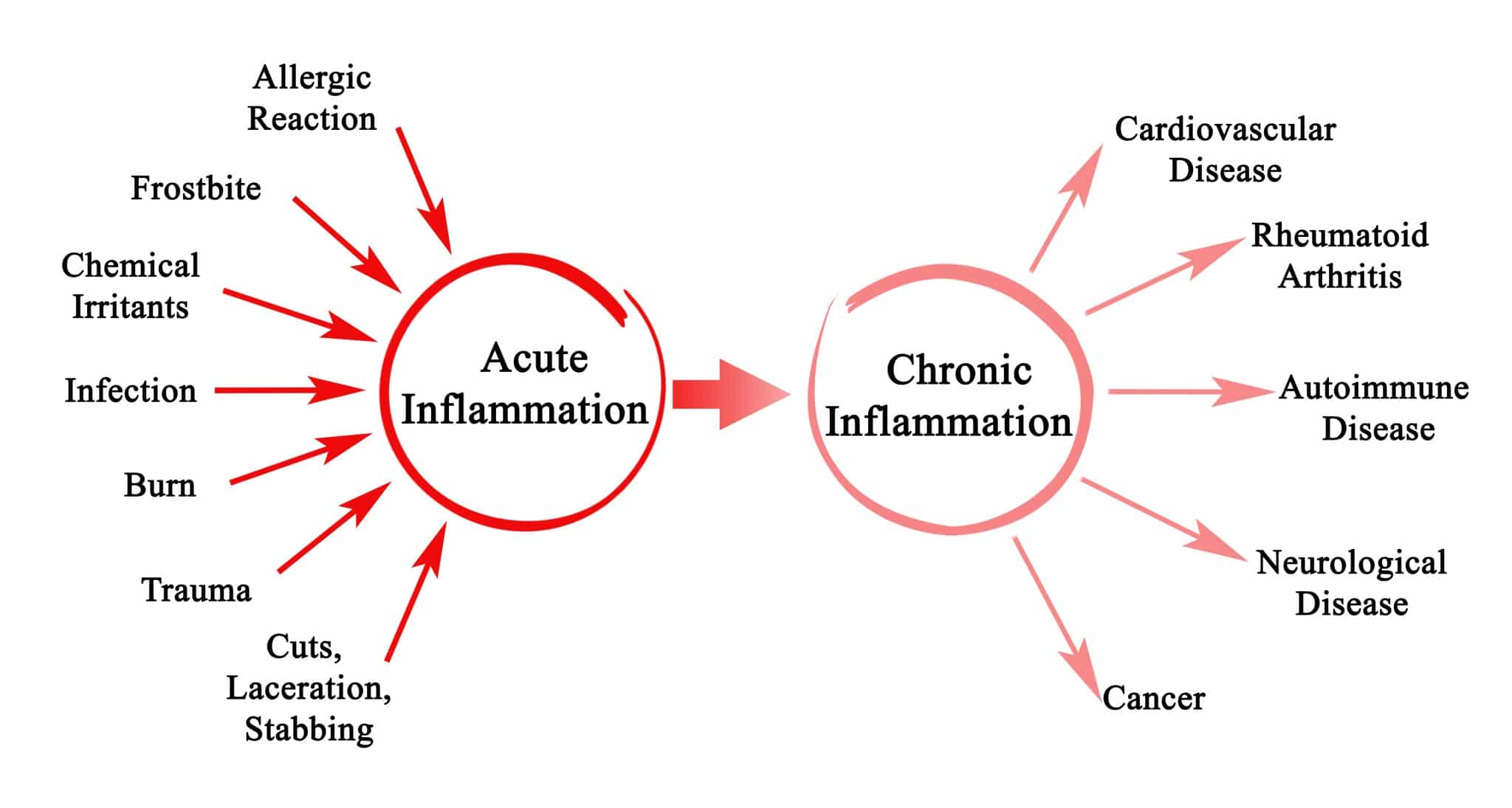
Digital Infrared Thermal Imaging (DITI) offers a revolution in aiding the early diagnosis of a wide range of diseases & dysfunctions – from rheumatoid arthritis to breast cancer, and frostbite to cardiovascular disease. In identifying acute and chronic inflammation – often the first signs of dysfunction – thermal imaging offers incredible opportunities to clinicians in the management of health & wellness.
The FDA approved the use of this incredible technology as an adjunct screening tool in 1982, which means that while it is not a standalone diagnostic tool, it has nevertheless been recognized as offering valuable additional insights into the screening process. The amazing fact about thermography is that it can identify possible areas of concern way before overt physical symptoms have appeared.
The sooner you identify inflammation, the earlier and easier it is to turn things around and support your health and wellness. Medical thermography is an amazing physical examination tool. ~ Studio Team
How Does Thermography Work?
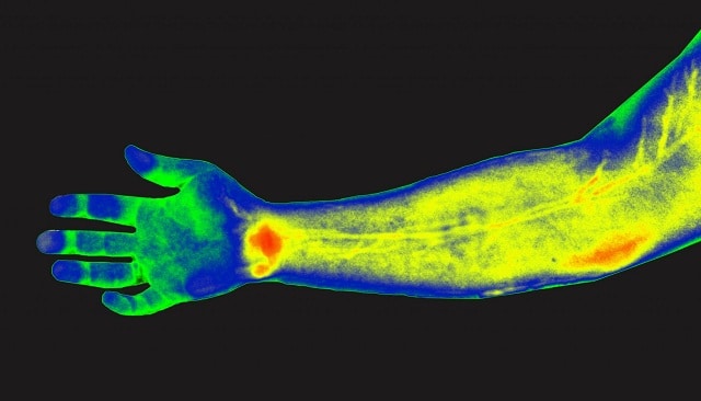
How does a thermal imaging scan work? Thermography, or thermal imaging, uses a heat-reactive camera to provide a highly effective, non-invasive, and dynamic measurement of the heat radiated from the superficial dermal microcirculation of our skin (approximately 1–2 mm below our epidermal surface) to pick up signs of inflammation, unusual patterns in heat or metabolic activity distribution, or asymmetry in heat (for example, both hands should show the same thermal heat pattern; if one does not, it might be an early indicator of arthritis in one hand).
Now, we know that the temperature and microcirculation of our skin are influenced significantly by inflammatory, metabolic, and toxic factors, so when we suffer the very early stages of degenerative disease (such as arthritis), we might not feel any physical symptoms, but physical damage has already, at this early stage, started to occur.
A thermal imaging scan will immediately pick up local changes in epidermal heat and blood flow, alerting us to the fact that something is happening so that we know that we need to run further tests. This is where the true value of thermal imaging lies – early detection before the structure of tissue has been altered (i.e., damaged) so that treatment options are as effective as they can be.
The Doctors Studio Mantra…. FIND IT AND FIX IT BEFORE IT BECOMES PROBLEMATIC!
Breast Cancer
A lot of thermal imaging research revolves around the value of DITI in breast cancer scans. The growth of a tumor requires angiogenesis (new blood cell generation), greater metabolic activity (due to nutrient and growth needs), and greater blood flow (enhanced vascular density).
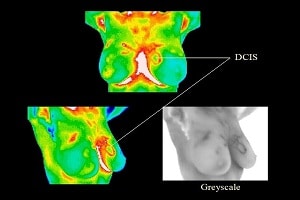
This adaptive (but unwanted) physiological activity would push the body out of its delicate thermal homeostasis (i.e., balance) in the very localized area where the tumor is growing. Thermal imaging can rapidly pick up on this unusual temperature & metabolic activity, and also clearly highlight the asymmetry between a healthy breast and a potentially cancerous one (i.e., where unusual thermal activity has been picked up).
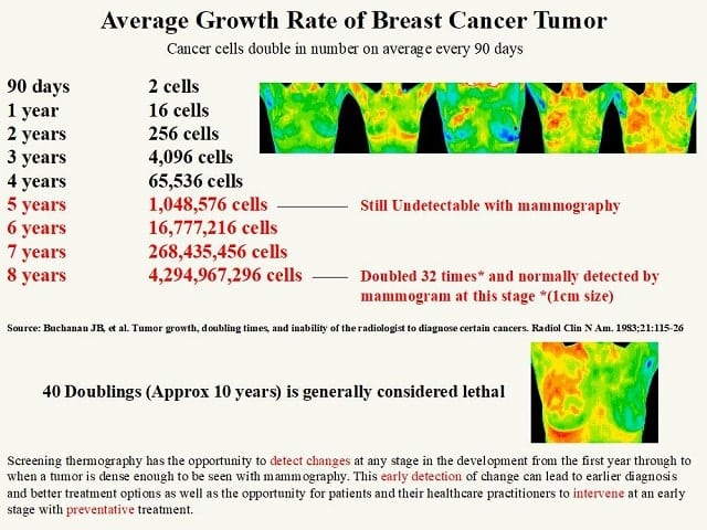
Together, this data immediately alerts a clinician to the fact that something anomalous is happening in that area of your body. If cancerous, the thermal imaging scan allows detection of potential carcinogenic activity way before a mammogram scan would routinely be used (mammograms – whilst the gold standard of breast cancer detection – require the physical presence of a tumor for a diagnosis to be made, by which point the disease may have significantly progressed).
The identification of that something could save lives, vastly increasing the patients’ survival outcomes & quality of life, decreasing the likelihood of cancer metastasizing, and significantly lowering the need for more aggressive later-stage treatments (such as chemotherapy).
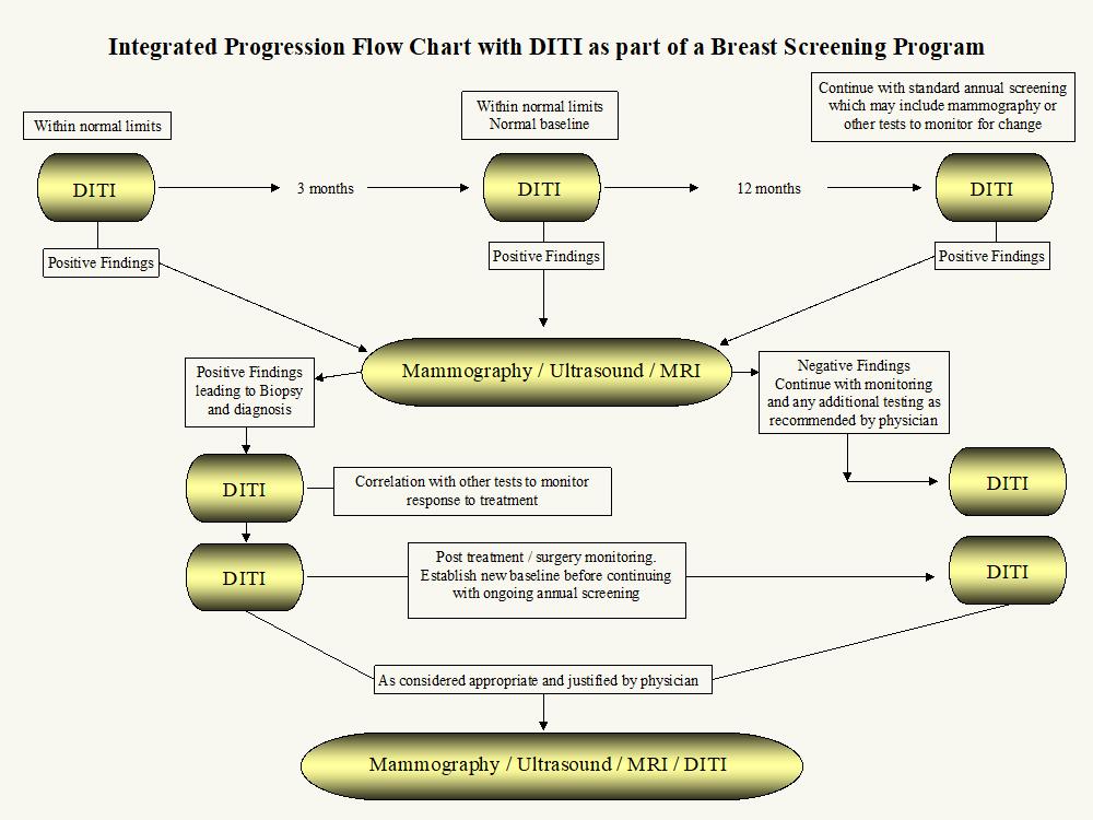
DITI’s function, ultimately, is to alert us – and a patient – to adaptations in the body that might require further investigation. In doing so, it provides a means of early detection of cellular changes with potentially life-altering outcomes. A brief review of the diagram below makes it clear that DITI can offer an extremely valuable ‘step 1’ in the breast cancer screening & diagnostic process.
Full Body Thermography
Thermal imaging offers an excellent preventative tool that can alert us to a vast range of abnormalities that may be signs of disease. Because the disease causes inflammation, and inflammation causes adaptations to thermal activity, DITI can offer a great means of raising awareness at an extremely early stage of a huge range of potential dysfunction.
It also offers incredible opportunities in the early and pre-emptive identification of musculoskeletal imbalances that can lead to injury, in the pinpointing of pain and inflammation, and in the management of chronic pain conditions.
How can DITI help with pain management?
Well, when blood flow is obstructed and decreased (due to an injury, or disease – or something as minor as sleeping in an odd position one night!), it can cause hypersensitivity of the surrounding tissue. If not immediately rectified, this hypersensitivity can lead to pain (and even eventual tissue death if the blood flow is not restored!). Returning blood flow to normal can eliminate pain altogether, so understanding the causes of hypersensitivity and disrupted blood flow remains key.
DITI also offers an excellent full-body scan for the presence of arthritis. Consider this; once you begin to experience pain, discomfort, or swelling, arthritic inflammation and joint damage may have already occurred. Diagnosing arthritis using IRT – before the painful accumulation of fluid, inflammation, increase in blood flow, swelling, heat, redness, and pain are experienced – offers a powerful opportunity to minimize damage caused by the disease, so that the patient receives the best prognosis possible. Subsequently, anyone, but particularly high-risk groups (i.e., women with a family history of arthritis) would benefit from the use of IRT in early, routine arthritis screening, offering an opportunity to treat symptoms before they manifest physically.
Ultimately, as with breast cancer, the earlier arthritis is picked up, the earlier it can be treated. The quicker the disease is halted in its tracks, the less damage it can cause to joints, leading to less pain, and greater sustained mobility, in the patient.
Head and Neck Thermography
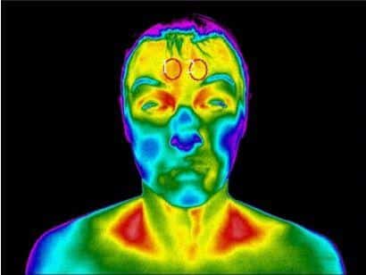
One specific area of the body for which thermography – a test that relies on heat – has proven powerful is in the head & neck region, partially because ‘the human body is considered to be a stairway of heat with the highest temperature in the forehead and cervical regions’.
For example, a 2014 study of thermography as a means of detecting CVD (cardiovascular disease) in India confirmed that blood pressure asymmetry, especially in the upper extremities (including head and neck) constitutes a clear, common early symptom in atherosclerosis, and that thermal imaging offered a clear ability to detect this.
An infrared thermal imaging scan might, subsequently, make the difference between a highly treatable CVD diagnosis caught at an early stage, and an untreatable stroke.
As such, thermography remains an excellent screening tool for head & neck-located dysfunctions, inflammation & disease. Head & neck-related uses include screening for cerebrovascular disease, sinus & facial inflammation, oral inflammation, thyroid dysfunction, periodontal disease, face & mouth cancer (including cervical lymph node metastasis and oral cancer) has emerged with strength in recent years.
Overall BENEFITS of Thermal Imaging (DITI)
There are many benefits associated with the use of this scientifically backed, progressive, screening tool. These include:
- Identification of areas of inflammation earlier & more reliably than other imaging devices.
- It is non-invasive, non-contact, and painless. This might be of particular value to women undertaking breast screening, as it does not involve often painful compression of the breast (NB. it should never replace a mammogram, however).
- The fact that it is non-invasive & painless.
- It is fast and relatively inexpensive.
- It does not involve any exposure whatsoever to radiation, and people can use it safely over time.
- DITI enables an effective treatment management tool for existing conditions, telling us how well your body is reacting to treatments, therapies, etc.
- It allows us to locate areas most viable for musculoskeletal injections.
- It offers an extremely valuable adjunct diagnostic tool, as approved by the FDA.
- It rapidly picks up on tissue asymmetry, which is a valid indicator of potential inflammation & dysfunction.
- It can detect inflammatory changes in breasts with dense tissue and implants.
- Hormonal and menstrual changes do not affect results.
- Medical Thermography is an ideal preventive medicine tool.
- It is safe for men, women, and children.
Is It Backed by Science?
Yes. There are over 800 articles in peer-reviewed journals referencing the utility of medical thermography as an adjunct diagnostic biomarker, with studies and research either previously undertaken, or presently underway, at universities and laboratories across the globe. The scientifically significant, predictive outcomes of DITI are well-discussed in academic literature, underscoring the medical value of DITI. At the Doctors Studio, we are led by medical science, and make clear the excellent adjunctive diagnostic value of thermal imaging.
As such, we offer thermal imaging as just one of a range of diagnostic & screening tools. Some private health centers offer DITI as a diagnostic tool in its’ own right, but this reflects a lack of medical knowledge; you can trust us to analyze your DITI results in the context of your medical history, and alongside the use of other tools to maximize your health & wellness outcomes.
Harness the Power of Thermal Imaging Today!
Here at the Doctors Studio, we consider thermal imaging to be one of the best choices available today in the achievement of preventative care, the ongoing monitoring of treatment, and the early detection of a range of potentially life-altering diseases & illnesses.
A thermal imaging scan is quick enough to fit into your lunch hour, non-invasive & painless!- so if you or someone you love would benefit from a thermography scan, contact us today. we would love to help you!
You can not fix a problem you don’t understand. Advanced diagnostics if often necessary to get at the root cause. Once the root cause is identified, the solution becomes possible.
Get to the Root of Your Health Issues At Last
Related Blogs
The Studio Method: Unraveling the Root Cause for Optimal Wellness
The Lifelong Athlete: Longevity Strategies for Peak Performance. Learn about cutting-edge interventions from Doctors Studio in Boca Raton.
Join the Wellness Revolution: Membership Benefits at Doctors Studio
Explore Doctors Studio's unparalleled membership benefits in Boca Raton—top professionals, exclusive discounts, holistic services for a wellness journey
The Five Root Causes Of Insomnia
What are the five root causes of insomnia? Find the root cause of your insomnia and get a full night's sleep—naturally.
Enlarged Prostate In Boca Raton: Treatment And Advice
How to find lasting treatment and genuine empowerment for an enlarged prostate in Boca Raton.
Should You Use Functional Medicine Telehealth Services?
Have you wondered if functional medicine via telehealth is a good fit for you? Learn more today!
What are Symptoms of Non-Celiac Gluten Intolerance?
An undiagnosed gluten intolerance can be a miserable experience.
Pro-Active Health: Is Mold Causing Inflammation?
Is mold causing inflammation? Florida is notorious for mold problems; here’s what you need to know to take charge of your health.
How to Find a Great Functional Medicine Doctor in Boca Raton
Functional medicine is a patient-focused, whole-body approach to healthcare. It is vastly different from the traditional approach that most of us are used to and centers around total wellness, preventative care, and treating or reversing chronic conditions.
Boost Immunity AND Burn Fat? LIPO-MIC: The Gift that Keeps on Giving!
Boost immunity AND burn fat with the ultimate Santa's little helper this holiday season! Lipo-Mic - the gift that keeps on giving!
Is Non-Celiac Gluten Sensitivity Making You Ill?
What is Non-Celiac Gluten Sensitivity? And is it responsible for your digestive and health problems? Read on to find out!
How To Get Started

Choose an Assessment Plan
Start the process by determining your current
wellness status.

Schedule a Consultation
Meet with an expert practitioner to review the results of you assessment and discuss your customized treatment plan.

Begin Your Wellness Journey
It's time to get back to balance and experience optimal wellness and quality of life

